After weeks of waiting the day finally arrived. It was Tuesday, July 23rd, 2013. Our destination for the morning was the Children's Hospital in downtown Detroit. We were scheduled to have a fetal echocardiogram at 9:15am. At this point we already knew that our baby had Down syndrome and there was a 50% chance he could have a heart defect. We were being optimistic and hoping for the best.
At 9:30am they called our name and we followed the sonographer into the room. I recall walking into the room and a chill came over my body. Whether the room was just cold, or I was beyond nervous is anyone's guess. Melissa didn't seem affected by it, but I was literally shaking. The sonographer quickly got started taking the various pictures. Baby Goulet was cooperating quite well and wasn't doing summersaults in the belly. The sonographer was able to capture the details of the heart much better than our normal ultrasound appointments. About an hour later the pediatric cardiologist was called in to look over the pictures and took some additional ones.
By 10:30am everything was done. Melissa and I were taken to a room where we sat down with the cardiologist. You could tell by the tone of her voice that something was wrong. She had two pamphlets in her hand and opened one up and started going over the diagnosis.
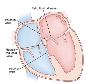
Baby boy has an atrioventricular canal defect. Basically the center of the heart has not developed - the area where the wall between the upper chambers joins the wall between the lower chambers. Also, the valves that normally separate the hearts upper and lower chambers arent formed as individual valves. Instead, a single large valve has formed that crosses the defect in the wall between the two sides of the heart. This defect can be repaired with surgery. During surgery, they will close the defect with one or two patches. Later the patch will become a permanent part of the heart as the hearts lining grows over it. They will also divide the single valve between the hearts upper and lower chambers and make two separate valves.
What we know today
When the baby is born there should be no immediate measures that need to happen. Baby should go home with us just like any other baby. He could appear to be more blueish then pinkish. The baby needs to do some growing before surgery should take place. Anywhere from 3 months up to a year, he will need to have open heart surgery. The baby could be in the hospital for up to 3 weeks, but due to their resiliency this is often less. The surgery will take place at Children's Hospital by Dr. Walters. He is the Chief of Cardiovascular Surgery and has done many of these surgeries. We have been told that the outcome is high for these types of defects. Because of enormous strides in medicine and technology, today most children born with this go on to lead productive lives. A child who has had this type of surgical repair will require life-long care by a cardiologist. Most children recover completely and won't need additional surgery. As you can imagine this news was devastating to us, but also hopeful at the same time. For now all we can do is wait and remain optimistic.
Atrioventricular Canal Defect
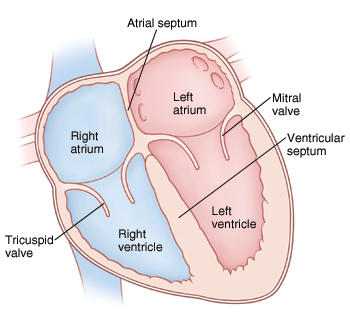
The heart is divided into four chambers. The two upper chambers are called atria and the two lower chambers are called ventricles. The heart contains four valves. The valves open and close to keep blood flowing forward through the heart. An atrioventricular (AV) canal defect is a large hole in the center of the heart. It's caused by the following problems with heart structure:
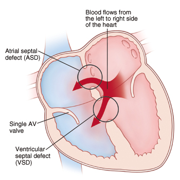
• Atrial septal defect (ASD): a hole in the dividing wall (atrial septum) that separates the atria in the heart.
• Ventricular septal defect (VSD): a hole in the dividing wall (ventricular septum) that separates the ventricles in the heart.
• A single atrioventricular (AV) valve: a single valve that develops in place of the separate tricuspid and mitral valves. These valves control the flow of blood from the atria to the ventricles.
Blood normally flows from chamber to chamber in one direction through the left and right sides of the heart. With an AV canal defect, blood flows through the ASD and VSD from the left side of the heart to the right side. This is called a left-to-right shunt. It causes more blood than normal to enter the right side of the heart. As a result, more blood than normal has to be pumped to the lungs. Over time, the extra blood flow causes the lungs to become filled with extra blood and fluid. This leads to a condition called congestive heart failure (CHF). Problems can also occur with the single AV valve. Because it's malformed, blood may leak backward from the ventricles to the atria (valve insufficiency or regurgitation). This causes the heart to work even harder.
Normal Heart

Heart with AVCD

Repaired AVCD

Click here for additional reading
At 9:30am they called our name and we followed the sonographer into the room. I recall walking into the room and a chill came over my body. Whether the room was just cold, or I was beyond nervous is anyone's guess. Melissa didn't seem affected by it, but I was literally shaking. The sonographer quickly got started taking the various pictures. Baby Goulet was cooperating quite well and wasn't doing summersaults in the belly. The sonographer was able to capture the details of the heart much better than our normal ultrasound appointments. About an hour later the pediatric cardiologist was called in to look over the pictures and took some additional ones.
By 10:30am everything was done. Melissa and I were taken to a room where we sat down with the cardiologist. You could tell by the tone of her voice that something was wrong. She had two pamphlets in her hand and opened one up and started going over the diagnosis.
Baby boy has an atrioventricular canal defect. Basically the center of the heart has not developed - the area where the wall between the upper chambers joins the wall between the lower chambers. Also, the valves that normally separate the hearts upper and lower chambers arent formed as individual valves. Instead, a single large valve has formed that crosses the defect in the wall between the two sides of the heart. This defect can be repaired with surgery. During surgery, they will close the defect with one or two patches. Later the patch will become a permanent part of the heart as the hearts lining grows over it. They will also divide the single valve between the hearts upper and lower chambers and make two separate valves.
What we know today
When the baby is born there should be no immediate measures that need to happen. Baby should go home with us just like any other baby. He could appear to be more blueish then pinkish. The baby needs to do some growing before surgery should take place. Anywhere from 3 months up to a year, he will need to have open heart surgery. The baby could be in the hospital for up to 3 weeks, but due to their resiliency this is often less. The surgery will take place at Children's Hospital by Dr. Walters. He is the Chief of Cardiovascular Surgery and has done many of these surgeries. We have been told that the outcome is high for these types of defects. Because of enormous strides in medicine and technology, today most children born with this go on to lead productive lives. A child who has had this type of surgical repair will require life-long care by a cardiologist. Most children recover completely and won't need additional surgery. As you can imagine this news was devastating to us, but also hopeful at the same time. For now all we can do is wait and remain optimistic.
Atrioventricular Canal Defect
The heart is divided into four chambers. The two upper chambers are called atria and the two lower chambers are called ventricles. The heart contains four valves. The valves open and close to keep blood flowing forward through the heart. An atrioventricular (AV) canal defect is a large hole in the center of the heart. It's caused by the following problems with heart structure:
• Atrial septal defect (ASD): a hole in the dividing wall (atrial septum) that separates the atria in the heart.
• Ventricular septal defect (VSD): a hole in the dividing wall (ventricular septum) that separates the ventricles in the heart.
• A single atrioventricular (AV) valve: a single valve that develops in place of the separate tricuspid and mitral valves. These valves control the flow of blood from the atria to the ventricles.
Blood normally flows from chamber to chamber in one direction through the left and right sides of the heart. With an AV canal defect, blood flows through the ASD and VSD from the left side of the heart to the right side. This is called a left-to-right shunt. It causes more blood than normal to enter the right side of the heart. As a result, more blood than normal has to be pumped to the lungs. Over time, the extra blood flow causes the lungs to become filled with extra blood and fluid. This leads to a condition called congestive heart failure (CHF). Problems can also occur with the single AV valve. Because it's malformed, blood may leak backward from the ventricles to the atria (valve insufficiency or regurgitation). This causes the heart to work even harder.
Normal Heart

Heart with AVCD

Repaired AVCD

Click here for additional reading

Leave a comment
Please login with your Facebook account if you wish to leave a comment.